


Optical lens expert explores microscopy
Carl Zeiss fascinating new division threatens to radically innovate the market for microscopic lenses


Major advances and progress in medicine and life sciences have always been spurred on by the latest developments in technology and the dedicated work of scientists. Like no other invention, the microscope has helped unveil the secrets of nature. With them, whole new worlds have become available, leading to numerous discoveries, without which we would be left with a limited understanding of everything around us.
The first scientific discovery based on microscopy dealt with the circulatory system and subsequently changed our view of the human body. Scientists have gone on to discover and explore life’s own building blocks, and recognised different types of bacteria and endeavoured against the spread of diseases such as cancer.
Once merely an ornament in the homes of the rich, the development of the conventional microscope at the end of the sixteenth century would lead to a great step forward for science, particularly in biology, and also in the development of materials and their usage; cast-iron and steel, minerals and polymers to name but a few. Largely considered to be the oldest existing optics manufacturer in the world, with lenses thought to be at the forefront of technological excellence; optical and precision mechanics specialist Carl Zeiss founded his optical workshop in the city of Jena in Germany in 1846. Carl Zeiss initially produced simple microscopes intended for dissecting work, and later, compound microscopes.
Before then, building a microscope was usually seen as a trial-and-error process, however his workshop was later to be considered an important hotbed for breakthrough concepts in optics and the collaboration between science and handcrafted technology.
Laying the foundations
In 1866, Zeiss met a young physicist called Ernst Abbe, and it was this partnership that led to the research of the theoretical principles of optical imaging in 1871, and the discovery of the Abbe-sine condition, which ultimately helped improve how well modern lenses could be made. A type of glass effective enough to fully test the theory didn’t exist at the time but a third collaboration with 30 year-old glass chemist Otto Schott produced a new type of glass, which could fully use the Abbe sine condition. This breakthrough – together with subsequent innovations of glass – enabled an entirely new class of microscope, the apochromatic lens, and enabled the large-scale production of high-powered specialist microscopes that allowed reliable, crisp imagery.
Helium ion microscopy allows researchers to drill holes in special substrates with extraordinarily small diameters for fast DNA sequencing
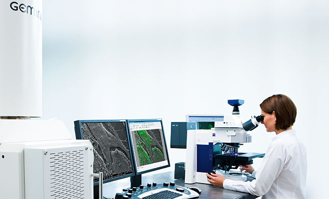
Today, Carl Zeiss Microscopy GmbH offers a uniquely broad portfolio: Innovative optic lenses and lighttrains in microscopes, high-performance confocal systems, and leading-edge fluorescence imaging instruments tailored to capture almost every photon emitted by a probe. Over seven different technologies exist which can optically slice living samples, even over long time periods. The resulting 3D images and movies enable reserachers to understand the basic dynamic principles of the metabolism of cells and a whole organism.The same holds true for the development of electron microscopy, right from the early steps in the late 1930s and after WWII, when the company started the development of Transmission Electron Microscopes (TEM) and later Scanning Electron Microscopes (SEM). There have been numerous research programmes into the treatments of tuberculosis, genome decoding, and the root cause analysis of cervical cancer, overseen by well-respected scientists, among whom a dozen are Nobel laureates who achieved their accolades with research done using the company’s microscopes.
At the heart of science
Last year, the optics powerhouse joined its light microscopy and electron microscopy units to form a single 2,500-strong business group. The combined division, Carl Zeiss Microscopy, is set to embark upon a new chapter in the history of microscopy – and aims to drive an industry trend which is seeing customers in both the research and industry fields regularly using both optical and electron microscopy systems, thus positioning the company as the only supplier of correlative microscopy systems.
Innovation at Carl Zeiss Microscopy was and is always driven alongside customers. Focusing to further develop strong partnerships with key researchers and scientists, Carl Zeiss Microscopy today employs a number of embedded scientists at high-profile facilities. These teams are currently working on projects of huge scientific significance, including several groups undertaking brain mapping, with the aim to build a connectome of the brain. Newly developed ZEISS scanning electron microscopes with built-in sample slicing capabilities are a main driver behind these research activities. With the extensive raft of knowledge acquired out of this cooperation, a highly-skilled service and support team can support Carl Zeiss customers to get their research jobs done efficiently. A global network of demonstration facilities and the ZEISS` Microscopy Lab in Munich supports the selection process of the best-suited technology by offering proof of concept support and the ability to collect images and data for grant applications.
Latest developments
In 2011, Carl Zeiss Microscopy won a coveted “Life Science Industry Award” which recognises best-in-class science suppliers. The company also won the 2011 “Best cell Biology Instruments (microscope-based) Award”, beating six other nominees in the category, and truly cementing its status as a cutting-edge research and production facility. Over the years the prestigious “R&D 100” award as well as the “Red Dot Design” award was awarded to microscopes and microscope systems from Carl Zeiss several times.
But there are already efforts under way to further advance microscopy. There are still vast areas in science, where knowledge is still limited and major efforts are needed to develop cures and new technological solutions. Most degenerative diseases such as Alzheimers cannot be treated; in material sciences the storage of electricity in batteries is still not sufficient performant to enable electro mobility.
To better understand processes on a subcellular level in cancer research, super-resolution micrsocopy is a major step forward. With over 50 research facilities now using the ZEISS ELYRA system, specific markers of the illness can now be better localised and observed in living cells. Bridging this functional perspective with structural results of electron microscopes opens a totally new world. For the first time the overlay created by the shuttle and find tool enables reconstruction of the neuronal network in the brain.
In geosciences, scientists are analysing shale-rock permeability by using high-end focused ion beams and scanning electron microscopes made by the company. The aim is to find natural gas and oil reservoirs and to decide whether it’s possible to extract oil and/or gas in a cost-effective, economical manner.
The latest projects underway include the unique ORION heliumion microscope – a technology that enables ground-breaking research in imaging, nanomodification and nanoengineering applications. For example, helium ion microscopy allows researchers to drill holes in special substrates with extraordinarily small diameters for fast DNA sequencing. This will in the future dramatically cut down the costs for individual sequencing, enabling personalised cures aiming at specific tumors or other diseases. With an unrivalled number of results and insights into the properties and characteristics of their research, it seems Carl Zeiss Microscopy will remain a global leader in driving science for years to come.