


JenLab offers a new quality check for corneal transplantation
Corneal transplantation is fraught with difficulties, particularly surrounding the evaluation of donated corneas. JenLab’s multiphoton tomography system aims to improve the odds for patients


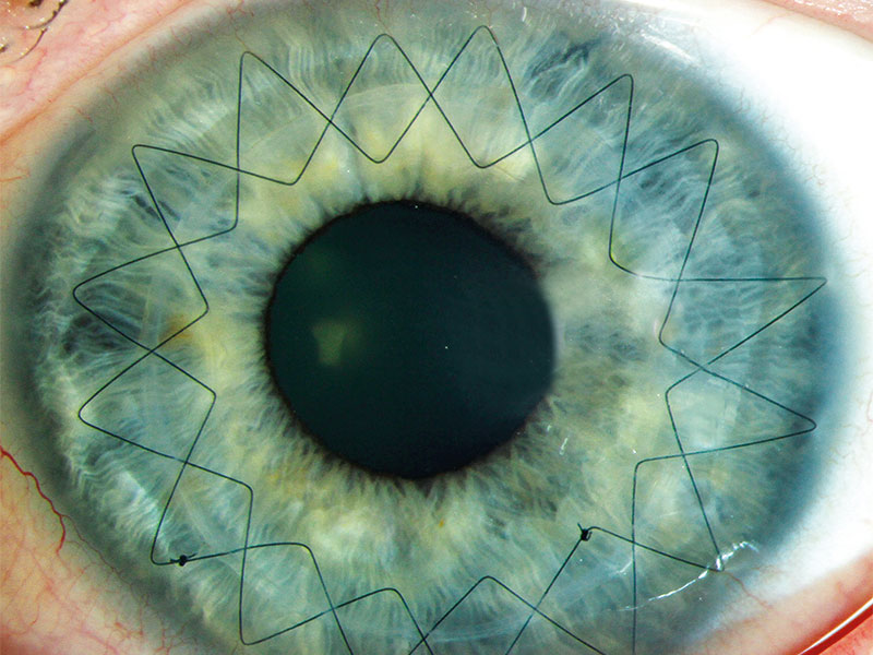
Following transplantation, the two star-like fissures seen here remain visible for as long as several months
Corneal diseases are the second biggest cause of blindness worldwide. When conventional treatments fail, a patient’s sight can only be restored through the transplantation of donated human corneas.
Corneal transplantation is the most common transplantation procedure in the world. However, there are not enough donors to satisfy demand, with the waiting time as long as nine months for a donated cornea. Adding to the problem, a large number of donated samples are unsuitable for transplantation due to the donor’s medical record or due to poor tissue quality, such as a low endothelium cell density. Normally, donated corneas are kept for days, or even weeks, in special containers prior to transplantation.
Corneal transplantation is the most common transplantation procedure in the world
Multiphoton tomography
Currently, quality checks on donated corneas are performed by slit-lamp examination, providing information on the corneal anatomy and morphology. At JenLab, we have developed a novel method with better resolution, offering additional information on cell metabolism, as well as on the structural organisation of stromal collagen. The method is called multiphoton tomography (MPT).
MPT based on near-infrared femtosecond laser technology is already in clinical use to provide non-invasive, label-free optical biopsies of human skin. Current applications include early detection of black skin cancer, evaluation of anti-ageing cosmetics, and measurement of skin thinning in astronauts after long-term space travel.
Now, with funding from the EU’s Horizon 2020 programme (grant 726666, LASER-HISTO, phase 2-SME), JenLab is pioneering the use of MPT for the improved quality check of cornea transplants, using optical metabolic imaging, collagen imaging, and lipid/water imaging. The metabolic state can be assessed through the fluorescence lifetime of autofluorescent coenzymes. The network of collagen fibrils can be imaged via second harmonic generation (SHG), where the infrared laser beam is transferred into weak blue light. Chemical information on the distribution of lipids and water content can be retrieved by rapid Raman (CARS) spectroscopy. CARS also opens a path to studying the pharmacokinetics of eye drops.
Clear view into the cornea
In JenLab’s MTP process, multiple detectors allow the simultaneous measurement of autofluorescence, fluorescence lifetime, SHG and CARS with single photon sensitivity. A typical optical section consisting of 512 x 512 pixels takes just six seconds. The intracellular cells and the collagen network can be seen immediately on the screen.
In a first clinical study at Saarland University Medical Centre, we demonstrated MPT images can be used to characterise the morphology of corneal layers, intratissue cells, the collagen network, and the metabolism of human corneas. Furthermore, a bacterial contamination can be detected.
Interestingly, the metabolism of a variety of intratissue cells has been found to increase with time after donation, whereas the metabolism of the important endothelial cells decreases. Thus, MPT can be used to optimise the storage procedure and to evaluate the right time for transplantation.
We believe that, within the next few years, MPT may be used for high-level quality checks of donated corneas for transplantation in order to increase the number of positive surgical outcomes. Furthermore, in vivo MPT will also very likely be employed on the human eye to diagnose diseases such as diabetes through a direct view into the body via the human transparent window: the cornea.