


NordicNeuroLab transforms fMRI technology
NordicNeuroLab is the winner of our 2015 award for Most Innovative Neuro-Imaging Solutions Provider. Yanlu Wang, an application specialist at the company, explains what it did to earn it


The NNL VisualSystem, NNL AudioSystem and ResponseGrip in use. NordicNeuroLab has been developing fMRI technology
Human curiosity knows no bounds, but the traditional means of satisfying it is usually self-defeating; all too familiar is the feeling of despair when we pick something apart to figure out how it works, only to find we are unable to put it back together again in proper working order. With age and experience, we learn to avoid this unhelpful behaviour, but are left unable to answer our curiosity about the world around us.
The invention of diagnostic imaging is arguably one of the biggest favours mankind did itself in the past century; medical imaging techniques allow us to visualise the human body without damaging it in the process. With recent advances in functional imaging, we can now not only visualise what the human body looks like, but also how it functions.
Advanced medical imaging techniques do save lives
Who we are and what we do
NordicNeuroLab is based in Bergen on the Atlantic coast of Norway. Originally a spin-off company from the local Haukeland University Hospital, NordicNeuroLab was founded in 2001 to design and produce equipment for functional MRI (fMRI): an advanced MRI application for the visualisation of brain functions.
Our main goal then as now was to make fMRI easy. Like many advanced MRI applications, an fMRI scan requires an intricate system consisting of many parts in addition to the MRI scanner itself. Because of this, only the largest institutions in the world were capable of providing the support needed for fMRI scans when NordicNeuroLab was founded – it was certainly the case in Bergen.
Known for its beautiful landscape and rainy weather, Bergen is also the home of Haukeland University Hospital, one of the first institutions in the world to conduct fMRI experiments. The leaders of the Bergen fMRI Group, and the founders of our company, decided to use their expertise and knowledge to provide fMRI systems in a bundled package. The system was designed so their peers and colleagues in other institutions could easily start their own fMRI studies, and thus NordicNeuroLab was born. Our fMRI system consists of all the parts (besides the MRI scanner itself) needed to perform fMRI scans. It is simple to operate, robust, and is made in Norway with a high Scandinavian standard of quality.
Over the past 14 years, we at NordicNeuroLab have kept our focus on MRI but also developed our product line-up to include hardware peripherals for patient comfort and interventional MRI applications, as well as software for processing advanced MRI neuro-applications. NordicNeuroLab products have been installed and are in use in 65 countries around the world.
The forefront of medical imaging
Over the past decade, fMRI has grown from a neuroscientific curiosity to a validated imaging technique with diverse clinical applications. One of the main goals of modern neurosurgery is to maximise quality of life, and it is often prioritised over life expectancy. Pre-surgical planning based on functional information allows neurosurgeons to plan surgery to achieve the best possible quality of life afterwards. FMRI can also be used post-trauma to locate functional deficits and optimise treatment plans.
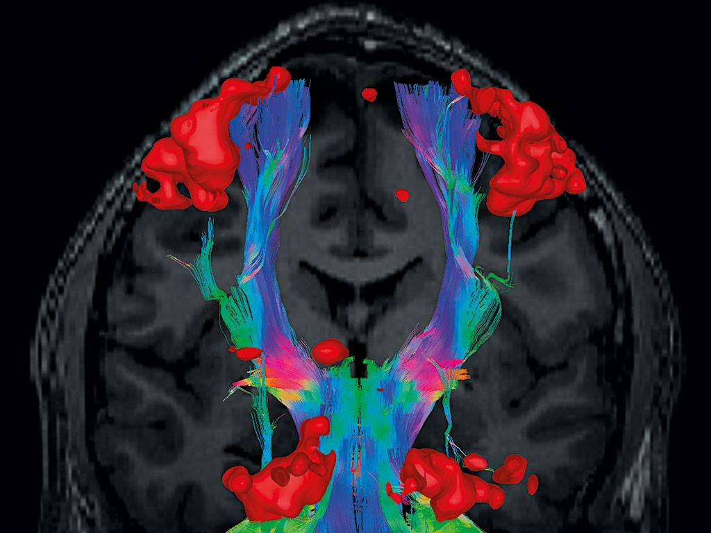
As new applications for fMRI emerge, we at NordicNeuroLab swiftly adapt our systems to meet their general needs, allowing hospitals and research institutes to focus on what they do best rather than spending resources setting up peripheral equipment and technical support. At the same time, we have developed support for applications that are traditionally symbiotic with fMRI, such as white-matter fibre tractography, MR perfusion scans, and hardware to improve patient comfort, as well as all real-time feedback inside the MRI scanner room to facilitate MRI-guided interventions.
At NordicNeuroLab we strive to bridge the gap between what researchers develop at the highest level of general development in neuroimaging, and what is applied day to day in hospitals and clinics worldwide. We believe advanced medical imaging techniques do save lives and improve the quality of life for people far beyond the image.
On one side, we have close working relationships with some of the world’s leading neuroimaging institutes and have developed software that is easy to use yet powerful in features. With our recent release of advanced MR perfusion post-processing software, research institutes and hospitals alike have access to tools that let them calculate how blood flows through the brain. The software enables institutes without prior support infrastructure to conduct MRI perfusion studies at the highest level of research and development.
On the other side, we are working with top industry partners to push advanced MRI neuro-applications to the next level in their clinical applications. Our clinical software package is designed to push potentially life-changing information to neuro-navigation systems, allowing neurosurgeons to plan neurosurgery and biopsies beforehand with more information than ever.
Traumatic brain injury can be detrimental to one’s well-being and quality of life, especially for those involved in professional contact sport. We work closely with some of the world’s leading neurocognitive institutions and veteran affiliated hospitals in the US to optimise treatment schemes for those who need them most. FMRI scans can be combined with traditional morphological MRI scans to visualise and quantify how brain injury affects one’s neurocognitive function; rehabilitation schemes can thus be tailored according to individual needs.
Electronics in general do not work well alongside an MRI scanner due to the extreme magnetic fields. With our high-quality, MRI-compatible displays, we are able to provide visual information with medical-grade clarity inside the MRI scanner room without interfering with the scanner. This allows MRI-guided interventions that take advantage of the flexibility and high quality of MRI scans.
We are constantly searching for the next big thing, but at the same time we are firmly rooted in our traditions. We believe MRI’s flexibility puts it at the forefront of the medical imaging field. Time and again, MRI has created stunning and beautiful images of our brain and body beyond what we could have hoped for, imaging aspects of ourselves we previously thought hidden. By unveiling the inner workings of ourselves, we will improve the lives of many, one small step at a time.