The New Economy transforming markets Series showcases the most exciting work in the tech world. Here, we look at what Ingenuity Lab and its partners have been doing to push forward healthcare
You’ve got to look after your health, and never was that truer than in these days of soaring populations in developing nations and rapidly ageing populations in developed ones In this supplement, we look at the work being done by Alberta-based Ingenuity Lab and its partners to combat threats to global health, from diabetes to cataracts
Type 1 diabetics currently rely on lifelong insulin injections to manage their condition, but research is underway on a transplant that could make treatment far less painful
Diabetes Helping the body help itself

While manageable, the process of daily insulin injections for type 1 diabetics is an inconvenience to say the least. The need to monitor insulin levels with frequent, inconvenient tests is the only option available to a number of people, impacting on freedoms and quality of life. This could soon change though, thanks to an innovative solution currently in development.

Research being undertaken with the assistance of Ingenuity Lab in the field of pancreatic islets transplantation could lead to type 1 diabetics having to worry less about their condition. Between the use of an alternative cell source to humans and the development of a scaffold that will support the growth of transplanted cells, implanting islets could finally become a practical solution.

Type 1 diabetes occurs when the pancreas, a small gland located behind the stomach, fails to produce the correct level of insulin a person needs. Insulin is a hormone that maintains blood-glucose levels in the body, and subsequently energy levels. If the body is failing to manage its energy levels correctly, it leads to a range of short-term problems, such as tiredness and weight loss. Long term, the effects are far more damaging, with potential issues ranging from kidney disease, heart disease, blindness and possible limb amputation. Apart from injections of insulin to keep levels correct, alternative treatments are often underdeveloped: an insulin pump via which the hormone can be administered, for example, is only practical for a relatively small number of patients. Another therapy alternative has been the transplantation of pancreatic islets. This solution might seem like an obvious one, but logistically it has always been impractical; as with any transplant therapy, there is a constant shortage of donors. Public awareness and education programmes can only go so far. However, a new source of cells is being investigated.

Continuing legacy
Dr Gregory Korbutt at the University of Alberta is carrying out research on an alternative source of islets, as well as a new way to implant them.

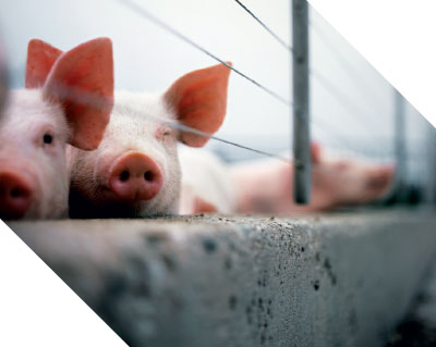
“One of the things we’ve developed here is using pigs as the source of insulin-producing cells”, said Korbutt. Pigs are a common candidate for human transplantation thanks to their availability, comparably sized organs and their genetic distance from humans. The work carries on the legacy of groundbreaking pancreatic research conducted at the University of Alberta. The school was the first to develop a standard for the implantation of islets, known internationally as the Edmonton Protocol, in a paper published in 2000. Korbutt, who was part of that original research team, said that, while the initial protocol recommended islets be transplanted into the liver, new research is now indicating there could be a better location. Along with the cell source, Dr Korbutt is also currently working on finding a better way to implant the cells.

ONE OF THE THINGS WE'VE DEVELOPED HERE IS USING PIGS AS THE SOURCE

“Then we are combining it with the technology from Ingenuity Lab”, he explained. “They’re able to make biomaterials. Ultimately, it would be a very non-invasive procedure if we were able to develop a transplant site – let’s say underneath the skin of patients. And that’s where Ingenuity Lab with their biomaterials are very useful.”

The islet transplantation doesn’t represent a cure for type 1 diabetes, but it could make living with the condition a far more manageable experience.

“It’s changing one form of therapy for another”, said Korbutt. “Instead of taking daily insulin injections, you’re getting a transplant. Seeing as it is a transplant, patients have to take anti-rejection drugs. But that’s where these biomaterials come into play.”

The research theorises that the use of a scaffold could create a space within the body that would receive enough blood flow to encourage transplanted pancreatic islets to graft onto their new host, survive and then begin producing insulin.

This is a challenge in two halves. The first is the source of pancreatic islets, which Korbutt is working on, but the second is the scaffold. This is the key to encouraging the cell growth that is needed for the transplanted cells to survive for any worthwhile amount of time. This is the portion of the project Dr Sinoj Abraham from Ingenuity Lab is working on.

New materials
Abraham said, when it comes down to it, the material of the scaffold makes all the difference. “The biomaterials we are making basically mimic the growth factors in the body, and then we also make the polymers with the different combinations. We customise the polymers so that we could easily customise it depending upon the patient, depending on the degradability, and all those things.”

What has made this innovation possible is the development of new materials that have a biological function built into them. Scaffolds used to be basically just a frame, with the patient's body having to develop its own growth factors. New materials can mimic the growth factors of the human body, so the process for the transplant integrating with the patient’s body becomes faster.

“We’re going to develop strategies where we can modify these biomaterials by putting molecules on that will help prevent rejection, and then the patients wouldn’t have to take lifelong anti-rejection drugs”, said Korbutt.

Pancreatic research is a competitive field to be working in, with several different strategies to solving the same problem currently in the works. One such project is the artificial pancreas, an electronic device that remotely checks glucose levels through a sensor placed under the skin. It wirelessly transmits information to a monitor worn on the body, and then transmits that information to a pump that maintains insulin levels.

Korbutt said, while the research is exciting, the technology isn’t quite there yet. “The problem with the artificial pancreas is the technology is not there to have a sensor that can detect patients' blood sugar and to release the insulin.”

The research partnership between Korbutt and Ingenuity Lab is still in its early days, but the collaboration has already found some traction with outside parties. “We established this collaboration I would say approximately nine months, so I would say that we are just starting”, said Korbutt. “We have just recently got funding from the Juvenile Diabetes Research Foundation, which is an international organisation that funds research into type 1 diabetes.”

Despite the challenges the research presents, Korbutt said he is confident the challenges can be overcome. “It’s research, so there’s always going to be barriers, but in research you do the experiment, if they don’t work you modify them and then you proceed. I think that a novel thing that we have here is we’re combining the biology with the biomaterial engineering expertise.

“In research, one group cannot solve all the problems, and that’s, I think, the novel part of this: we’re combining technologies. It strengthens the project and I feel will improve the probability of succeeding.”

While much about the brain is still unknown, Ingenuity Lab is working on a tool that could help us better map its pathways and revolutionise neurology as we know it
Neurology Mapping the human brain

In spite of the countless leaps and bounds that have been made in medicine, there is still a great deal we do not know about the human brain: how it functions in real time; what causes disruptions; and exactly where those disruptions take place. Finding ways to reveal such fundamentals is crucial in establishing a greater understanding about numerous diseases that affect millions each year. This knowledge holds the key to developing more effective and efficient treatments; it could even make the prevention of many issues a distinct possibility.

That better understanding of the brain could be here sooner than we think. At the cutting edge of such research is the team at Ingenuity Lab, whose focus on neural probes could well solve our unanswered questions. The institution’s programme is finding new ways to leverage the tools of micro- and nanofabrication, together with nanotechnology, in order to develop enhanced devices that can accurately interface with the brain.

The hope is these devices will improve our understanding of how the brain works, which, in turn, will aid disease diagnosis and treatment. Specifically, the systems Ingenuity Lab is developing include microfabricated electrocorticography (ECoG) and biochemical transistors, which can precisely measure and map the electrochemical signalling molecules of the brain.

Brain freeze
The biggest challenge in understanding the innermost workings of the brain is the temporal and spatial resolution of information. The brain is extraordinarily complex; the locations where activity occurs need to be mapped with incredible accuracy, but this is simply not possible at present.

“It’s like trying to find somebody’s house with Google Maps from a satellite dish 40,000 feet away; you can’t see the house until you zero down to a really fine dimensional space”, said Dr Carlo Montemagno, Director of Ingenuity Lab and a world-leading expert on nanotechnology. “Trying to understand how the brain works is very much analogous to that; if you can only get very coarse measurements of both position and timings of where things are occurring and when, it is hard to define exactly what causes disease, how things function, and how to then design the most appropriate treatments.”

We think that we’ll be able to do a better job of mapping function in the brain

ECoGs, which are electrodes that are placed on the surface of the brain, are used primarily for mapping brain function; they hold the key to Ingenuity Lab’s work. “By incorporating different specific signalling molecule receptors to make chemical transistors that respond to specific chemical signals, we’re able to measure electrical impulses, in addition to the chemical signalling that goes on in various portions of the brain. This allows us to see how signalling and electrical information changes, based upon position and time”, explained Montemagno.

Through the use of ECoG arrays and 3D printer technology, Ingenuity Lab is working on dimensionally designed probe systems for individual patients. This approach will give scientists the surface topology they need to give better context to existing probes on the brain. “Coupled with chemical measurements, we think we’ll be able to do a better job of mapping function in the brain – and with better signals and noise ratios too”, said Montemagno.

Such advanced neural probes will provide far more precise mapping for disorders such as epilepsy and Parkinson’s disease, aiding treatment immeasurably. Moreover, they will also enable deep brain stimulation probes to be placed more precisely than ever before, which could provide a far greater insight into cognitive processes, defects and patient progress. “The technology that we’re using does two things”, said Montemagno. “Firstly, it allows us to make probes that map the anatomical variation that occurs in the normal population, so that sensors are placed at the best location possible to get the signal information that you want. Secondly, this technology offers a much higher density of sensors on the probes, which allows us to get higher spatial embedded temporal resolution of what's actually going on.”

Light bulb moments
In addition to successfully making these innovative ECoG arrays using 3D printers, Ingenuity Lab has also developed new types of conducting polymer inks. “We have found a way to interface them to channel biological receptors, which are the chemical receptors that cells have for communication, and then incorporate them within our probe system in order to map everything together”, Montemagno continued. “So the basic building blocks are all there, and we’re now refining the manufacturing production process to get the precision, repeatability and resolution that we need, so that we can start translating it”.

Using soft materials, such as polymers, coupled with the latest manufacturing technologies, the development in terms of both cost and performance of the lab’s systems is monumental. “I saw an opportunity there through a medium-term research effort that could significantly improve the performance in the field, which we can push to the next stage with our project partners, Neuralynx”. Based in Bozeman, Montana, Neuralynx is one of the world’s largest suppliers of the data acquisition technology associated with both penetrating and surface-mounted probes. “For us, it was an obvious choice to work with them on developing these products, so that we now have a natural outlet for moving them to the marketplace.”

Of course, with any innovation there are challenges to overcome before a product can be deployed on a mass scale. For this project, the reliability of neural probes in terms of manufacturing, storage and performance needs to be assured before moving forward. “Doing a science experiment and demonstrating that something is feasible is one thing, translating it into a deployable technology is very different. This means ensuring that not only does it work, but that it works all the time and with the same level of performance. We also need to ensure that it can be produced at a price point that makes the product economically viable – so those are the targets that we are converging on right now”, Montemagno explained.

With few hurdles left to jump, the viability of this product’s commercialisation is near. In fact, Ingenuity Lab has plans for its prototype to be on the market within two years. That these highly precise neural probes will be produced at a lower price point than current counterparts, while also achieving a significant advance in both information captured and information actionable by investigators and physicians, is a monumental advance in neurological studies. With an enhanced understanding of the way the brain functions, researchers can design far superior therapies for a whole range of diseases, potentially saving the lives of people around the world. Indeed, with tools for precise brain function mapping in place, the lab’s upcoming offering could be nothing short of revolutionary.

Once considered useless tissue, the meniscus is actually critical to keeping knees working properly

Research is being undertaken to develop a solution to meniscus tears that doesn’t end in osteoarthritis
Meniscus Fixing up the joint

In February 2016, an injury forced tennis great Roger Federer to withdraw from two high-profile tournaments. While many thought he sustained an injury during his loss to Novak Djokivic at the Australian Open, Federer later revealed that was not the case. “I woke up, I don't know exactly remember what happened”, he said at a press conference. “I think I was going to run a bath for the girls. I made a very simple movement, turned back, heard a click in my knee. I went to the zoo. My leg was swollen.”

The injury was a torn meniscus, and the 17-time grand slam champion had to undergo arthroscopic surgery to fix the damage. Though common among professional sportspeople, a torn meniscus is an injury anyone could sustain from a careless step. Despite this, long-term treatments have been deeply flawed.

However, thanks to work being done with Ingenuity Lab, the way a torn meniscus is treated could soon be very different. With the development of 4D printing technology, the possibility of an artificial meniscus that can weather sustained punishment and is customised to an individual is nearing reality. Such an implant could prevent the need for a full knee reconstruction later in life.

Incredibly resilient
The meniscus is a wedge-shaped piece of fibrocartilage located between the thighbone and the shinbone, playing a critical role in the proper function of the knee. It stabilises the joint by distributing a person’s body weight across the whole shinbone while also lubricating the movement of the knee. These tiny pieces of cartilage are incredibly resilient; they support the entire weight of a person across a very small surface area.

Despite their strength, a tear is not an uncommon injury. Naturally sportspeople are particularly susceptible since they place a significant amount of stress on their knee, but a simple misstep for the average person could be all that is needed to do the damage. Because of the nature of the meniscus, treating a tear has always been a difficulty.

Dr Adetola Adesida did his PhD at the School of Pharmacy at the University of Manchester. He also studied at the School of Biological Sciences in Manchester and did his postdoctoral fellowship at the Wellcome Trust Centre for Cell-Matrix Research. But it was his one-year fellowship at the Harvard Medical School in Boston that brought him into the field of tissue engineering. He is currently working on a solution to torn menisci at the University of Alberta, and said the role of the meniscus has long been misunderstood.

Because of the nature of the meniscus, treating a tear has always been a difficulty

“The problem is that for many, many years surgeons would just take them out when there was a torn meniscus”, Adesida said. “It was regarded just like the appendix; that it was a useless piece of tissue.”

While removing a meniscus might temporarily repair the knee, it also allows cartilage to rub on cartilage within the joint. This isn’t an immediate problem, but the cartilage will inevitably wear out later in life. This leads to painful bone rubbing on bone and eventually early onset osteoarthritis. Young people who have a meniscus removed and develop osteoarthritis may have to wait years before they become eligible for a knee reconstruction.

These days, the function of the meniscus is better understood and surgeons now only remove the damaged parts when an injury is sustained, salvaging what they can. It can’t be stitched back together because it will inevitably rupture again. It also cannot heal, since two-thirds of the width of the meniscus is not exposed to blood flow. While preferable to completely removing the entire meniscus, Adesida said removing the damaged parts still leaves a lot to be desired.

“Now, if you compare two-thirds being taken out that is damaged, you only have one third left. One third is not able to replicate the natural function of the meniscus at all.” The solution Adesida has been working on with Ingenuity Lab is the development of a 3D-printed replacement.

Challenging materials
The struggle with developing an artificial meniscus is finding a material strong enough to endure the pressure the average human places on their knee, with many synthetic options simply not resilient enough to flex the way a meniscus does. Adesida is working on finding a way of combining a 3D biomaterial that will not only support the joint, but also incorporate stem cells that can stimulate the regeneration of the natural material of the meniscus.

“Then you can stimulate them to make the natural components of the meniscus itself, so you can basically replicate the mechanical properties of the tissue”, Adesida explained. He is working with Dr Anu Stella Mathews, a research associate at Ingenuity Lab with a PhD in chemical engineering. Whereas Adesida is working on the cell growth that will establish the development of the meniscus, Mathews is developing the material that will support the cell growth.

“In tissue engineering, there are many scaffolds they use which are FDA approved, so the major challenge Dr Adesida’s group gave us was to develop a material that the cells are happy with and we can print”, said Mathews. “Then we can at last implant the meniscus.”

What sets Ingenuity Lab’s scaffolds apart is the use of its patented 4D printing technology. While the term ‘4D printing’ can have many different meanings, the fourth dimension of Ingenuity Lab’s material is in what is added to the scaffold.

“In our case, we are inputting biological functions or bio membranes and proteins into polymers”, Mathews said. “Then we are finding the optimal composition of them and printing them so that the printed material will have a bio-function at the end.” Unsurprisingly, developing the material and cell source is not a simple task.

Future feasibility
“My lab really focuses on the cellular side of things, so we’re looking at trying to really identify the cell source, and by that I mean what cells will be ideal for actually making new meniscus tissue”, Adesida said. Stem cells can be sourced in many different ways, and finding the right ones for the job is a challenge. From the material side, Mathews said finding the right environment to promote cell growth is the biggest problem.

Another factor is keeping the cost of the procedure low enough so it is not completely unaffordable to the average person. Apart from the costs that go along with printing the tissue, another focus is keeping the amount of time an orthopaedic surgeon needs to work on a patient down to a minimum; too long, and the bills that would result from the time spent in surgery would make it impractical. While the project is still very early, Adesida said keeping factors like this in mind will make sure the solution they are working towards is a practical one. To people who have sustained a meniscus injury, a practical surgical solution could make a world of difference while also preventing the agonising onset of osteoarthritis.

Energy and resource efficiency is key for modern industry

Water usage, in particular, needs to be environmentally stable

Ingenuity Lab is developing new, low-cost separation technologies to purify water streams, promote recycling and protect the environment
ZIKA Protection through detection

In May 2015, a virus that up until then had been relatively unknown caught the world’s attention. Named after its site of origin, the Zika Forest in Uganda, the disease was first identified in humans in 1952; it is spread by a breed of mosquito called Aedes aegypti and was once thought to show few, innocuous symptoms. Similar to other flaviviral infections, such as dengue virus, the West Nile virus, yellow fever and tick-borne encephalitis virus, infection can cause fever, headaches, rash and joint-pain.

However, most adults who are infected do not experience any symptoms at all and can recover without any symptomatic treatment. Since the Zika outbreak in French Polynesia in 2014, however, there has been a significant increase in the number of patients who have been detected with Guillain–Barré syndrome (GBS) as well. GBS is often preceded by a bacterial or viral infection, such as Zika, yet can be far more serious as it weakens immunity and affects the peripheral nervous system.

Similarly, Brazil’s Zika outbreak in 2015 resulted in an increase in the number of microcephaly cases by around 2,700 percent. The congenital affliction is associated with deficient brain development in infants, which is often fatal within the first year. For those who survive to adulthood, they do so with severe physical and mental health problems, while the cost of medical care is astronomical.

As terrifying as this epidemic of microcephaly is, it may not be all that results from Zika. “The recent findings conclude the relationship between the Zika infection and neurological disorders, which might not be limited to GBS and microcephaly”, said Dr Sibel Cetinel, a leading nanotechnology expert at Alberta-based Ingenuity Lab. “Zika infection therefore creates a huge risk; greater than was even considered for both adults and unborn infants.”

Since there is no available treatment for the Zika infection, prevention is crucial

Rapid spread
According to the World Health Organisation’s August 2016 report, 68 countries have reported an outbreak of the Zika virus since early 2015. “Among these countries, 11 of them represent evidence of human-to-human transmission, rather than mosquito-borne transmission”, Cetinel explained. “The routes of human-to-human transmission are suggested to be sexual, blood transfusion and perinatal transmission from mother to foetus”. In fact, the rate at which the virus has spread across the Americas has been nothing short of astronomical, particularly when compared to that of, say, the West Nile virus.

In response to Zika’s looming threat, the US Centres for Disease Control and Prevention (CDC) has issued warnings to both pregnant women and those considering pregnancy against travel to over 50 countries in the Caribbean, South America, Central America and the Pacific Islands. Furthering concerns was the July 29 announcement of “active local transmission” in Wynwood, Florida – the first time the CDC has issued a warning against travel within the US since being launched in 1946. This was swiftly followed by another “active local transmission” warning for the Miami Beach area on August 19, leading Florida-based obstetricians to advise their patients against getting pregnant.

Since there is no available treatment for the Zika infection, prevention is crucial; limiting the transmission routes, particularly in terms of human-to-human transmission, can do a great deal to limit the spread. “In order to achieve this, rapid and wide screening of individuals is imperative, which can only be carried into effect with fast, specific and cost-effective detection systems”, said Cetinel. In a bid to implement such a system, Ingenuity Lab is using its advanced experience in various detection methods, as well as its expertise in surface chemistry and molecular interactions.

Detection methods
There are two broad approaches to Zika detection. The first is the general serological method, which is based on the detection of an antibody or viral antigen. “In the case of Zika, the serological approach lacks selectivity, due to the antigen similarities of related flaviviruses”, said Cetinel. “As such, this type of diagnosis may result in cross-reactivity for the patients who have been infected with other viruses, such as dengue.” As the second common approach is based on nucleic acid methods, it is able to provide selective detection. Unfortunately, however, due to the specific instrumentation and technical expertise required for this method, it comes with higher costs, as well as a longer diagnosis time.

This is where Ingenuity Lab comes in. “Our approach depends on the specific selectivity of Zika viral antigens, as we can use antigen specific antibodies and peptides for the detection”, explained Cetinel. “The surface of the sensor we have developed is decorated with these selective peptides, so that their specific binding interaction with the antigens » are recorded with the sensing platform, either with an indication of colour or signal change. The colorimetric method is designed to give qualitative data, which is either positive or negative. On the other hand, we have also designed more sensitive sensing platforms that utilise the same peptides in order to perform quantitative measurements”.

The main challenges of Zika detection methods, especially in the circumstances of an outbreak, are selectivity, cost-efficiency and point-of-care diagnosis. The method being developed by Ingenuity Lab, however, is designed to overcome them all. First of all, the selective peptides can specifically recognise Zika, but not other flaviviruses, such as dengue, eliminating cross-specificity reactions. Secondly, the cost is as low as that of glucose detection strips because the qualitative device utilises a paper-based platform, without any requirements for additional instrumentation. Finally, and of particular significance in terms of efficiency and mobility, is that point-of-care diagnosis is possible without any need for laboratory equipment or trained personnel.

In terms of the results already achieved for this project, Ingenuity Lab’s initial step involved first identifying Zika viral antigens. Both specific antibodies and peptides were then selected against Zika antigens on the basis that they would not interact with other flaviviruses – dengue fever in particular. The next stage in the project was to assemble the actual device, and then begin testing its efficiency as a diagnostic tool. “The final stage of testing the device will be the utilisation of patient serums. At that stage, the reproducibility and reliability of the system will be confirmed”, Cetinel explained.

Looking forward
Given the severity of the Zika epidemic, numerous research groups and drug companies around the world are working on a cure. They are focused on the development of a vaccine that can both control the endemic and eradicate Zika in the long run. Unfortunately, first phase I trials are only just being initiated; it will take quite some time for these drugs to come to market, and that will only happen if human clinical trials reveal their efficiency.

That said, there is still hope. What is most exciting about Ingenuity Lab’s work is its possibilities, not only for diagnosis, but also in terms of both treatment and protection. “Even though we initiated this project to create a more specific detection platform for Zika, the specific peptides and antibodies we have generated can find their application in targeted treatment, especially for the protection of embryos”, said Cetinel.

The lab’s work comes at a vital point: with local transmission having occurred in the US and given the extreme dangers faced by newborn babies, Zika poses an alarming threat that continues to worsen – and rapidly at that. The scientific community is therefore charged with a duty to find solutions fast; time is not a luxury they have. Ingenuity Lab, however, offers some optimism in this regard; the institution’s work on Zika detection could well be the start of a new phase in which we can actually control the damage caused by this terrifying virus.

Gene sequencing is moving beyond just reading genomes and is developing into a diagnostic tool, allowing treatment to be customised to a patient’s DNA
Genomics Boosting detection

A seemingly impossible task, scientists have calculated the approximate number of cells that make up the average human being: their count was a whopping 37.2 trillion. Knowing this spectacular figure, finding the comparatively tiny number of cells that make up a viral infection seems impossible. However, measurements this precise are now within the grasp of medical science, potentially resulting in highly customisable treatments.

While we have been able to sift through the most abundant examples of cells in the body, many secrets have been hidden in obscurity. If science is to continue unlocking the mysteries of the human body, a way to sort through the information it stores, even in the smallest amounts, is necessary.

Research being conducted by Ingenuity Lab in the field of genetic sequencing is improving the tools we use to find genetic material. While the development of next-generation sequencing (NGS) has allowed a greater amount of genetic material to be sifted through in a short period of time, the ability to identify microbes and viruses that only exist in minute quantities has only just begun to emerge. With improvements to current systems under development at Ingenuity Lab, these viruses could soon be easily identifiable – changing the way many conditions are treated.

The human genome
While recent developments have led to rapid growth in the field of genome research, its history dates back over 100 years. In 1871, Swiss physician and biologist Friedrich Miescher published a paper identifying his discovery and isolation of ‘nuclein’, a new molecule he discovered in the nucleus of cells. Nuclein would later be renamed deoxyribonucleic acid, or DNA, as its role in human development was gradually understood.

It took $2.7bn and 13 years for the first human genome to be sequenced: the cost can now be as low as a few thousand dollars

It took years before the real function of DNA was identified. In 1952, the Hershey-Chase experiments proved DNA carried the information living cells used for inheritance, now better known as genetic information. In this very early stage, the experiments proved that, when bacteriophages infected bacteria, they injected their DNA into the host and passed on their genetic qualities. While scientists had suspected this was the case for a long time, it hadn’t been proven until this point.

Along with appreciating the role DNA plays in human development, genome research has also led to the » understanding of genetic disorders. Mutations and errors in genes can have wide reaching and varied effects on the human body, including conditions such as cystic fibrosis and muscular dystrophy. When the role DNA plays in human lives became apparent, mapping the human genome to begin identifying which genes were responsible for what became a necessity.

The Human Genome Project began in 1988 with the US National Institutes of Health's director, James Wyngaarden, assembling scientists, administrators and policy experts to set the foundation for a study that would last 13 years. The next year, the National Centre for Human Genome Research was established, and work began on mapping the human genome. In 2001, the Human Genome Project published a 90 percent complete sequence of the three billion base pairs that make up the human genome. Altogether, it was found humans have approximately 20,000 to 25,000 genes.

The next generation
Dr Weiwei Wang is a research associate at the Department of Medicine, University of Alberta, Canada. He received intensive training in genomics, transcriptomics and proteomics, obtaining his PhD from Beijing Institute of Genomics, Chinese Academy of Sciences in 2008. In 2009, he decided to join Gane Wong's lab at the University of Alberta and to develop NGS-based assays for biomedical and clinical research. He said the way genes are analysed is changing.

“The DNA sequencing method is changing from low throughput and high cost, to high throughput and low cost; from the first generation based on the Sanger method of sequencing, to second and third generation as more and more nanotech gets involved”.

Since the Human Genome Project, a number of new and more efficient methods of reading genetic material have emerged. It took $2.7bn and 13 years for the first human genome to be sequenced: the cost can now be as low as a few thousand dollars. This increased speed and development has provided the opportunity for science to make breakthroughs in understanding genetic material that could have taken decades in the past.

NGS is something of a catchall term for the modern techniques used to read the genetic code of biological organisms: it has already been used to read the genomes of animals, plants, bacteria, viruses and other microorganisms at a far cheaper and quicker rate than was possible in 1988. This information has been used to develop a wide range of medical tests, including the early diagnosis of genetic abnormalities and the detection of microbial pathogens.

Beady little eyes
Beads are a crucial component in one of the modern ways genes are identified and sequenced. A sample of double-stranded DNA is taken and then broken up into fragments using restriction enzymes. Sequences of DNA, called adaptors, are attached to the fragments, and then resin beads are added to the mix. DNA sequences placed on the beads are complimentary to the sequences on the adaptors, allowing the fragments to bind to the beads. This isolates a DNA sequence to a bead, allowing it to be identified. It’s a far easier and faster process than the Sanger method used during the Human Genome Project.

While a significant improvement over previous methods, this more modern method of capturing DNA does have its limitations. One problem is its ability (or lack thereof) to target and detect low-abundant sequences: sequences of DNA that don’t have a large concentration within the body. While possible, the process costs an excessive amount of money, making it highly impractical and not suitable for widespread use. Seeing a need for improvement, Ingenuity Lab has been working on new technologies to find low-abundant sequences from clinical samples using probes to capture targets of approximately known content, such as viruses.

This has led Ingenuity Lab to develop InBeads, a more advanced version of the beads used in standard capture kits, to improve the way genetic material is scoured for information. Wang said the development of InBeads would lead to a less invasive, high performance diagnostic system for precise, personalised medicine.

“Direct NGS has limited recovery rate on low abundant target sequences, where the target reads less than one percent of the sample. Usually our targets, like infectious agents or tumour DNA, are around that low level in clinical samples, buried in greater than 99 percent human genomic background. InBeads can be used to enrich those targets to bring the signal high above the background.”

The results have so far been promising: relative to commercial capture kits, early tests have shown an improvement of the detection of low-abundant targets by a factor of between 100 times and 1,000 times.

Superparamagnetism
InBeads are spherical magnetic particles with a diameter of approximately 275nm. They are an improvement over other commercial beads on the market thanks to their larger surface area, stronger magnetic property, and unique surface functionalisation. The results of the changes Ingenuity Lab have made are much better performance in biomolecules capture and separation. Wang explained one of the nanotechnology principals implemented within InBeads is superparamagnetism.

The technology has the potential to improve the way new viruses are identified, characterised and associated with disease

“Superparamagnetism is a type of magnetism that occurs in sufficiently small magnetic nanoparticles”, he said. “Briefly, particles show relatively strong magnetic response when an external magnetic field is applied, but zero magnetic response when the external magnetic field is removed. Superparamagnetism allows particles to freely capture the ‘interested’ biomolecules in the solution, then effectively separate them by applying even a simple block magnet.”

The advancement of technology could make for a significant improvement in the way diseases are diagnosed. In an early demonstration, hepatitis C was identified much more efficiently than any of the traditional assays used in hospitals worldwide.

As well as diagnosis, the technology also has the potential to improve the way new viruses are identified, characterised and associated with disease. Additionally, it could be used for other healthcare applications including screening for early signs of cancer or inherited genes that could be associated with an increased risk for certain diseases. The technology also has potential applications outside human medicine.

Wang said there are still obstacles to overcome. “We will need to finish our tests on enrichment of DNA, RNA, and protein targets from mock samples with different abundance of human background, as well as certain scale of clinical samples in our good manufacturing practice labs and submit related patents, then we can move onto next step for commercial product.”

While developing the project, Wang explained limited clinical samples and the very low abundant targets buried in huge amounts of background noise have been notable challenges. There are also the difficulties that go along with making the system commercially viable; the whole process needs to be as fast and as cheap as is practical while still being effective. There is also the long-term stability of the beads, as well as maintaining consistent performance with large-scale preparation of them.

The longer-term prospects for the project could have a significant impact on the way people are diagnosed and treated. In the past, this was done based on averages and generalisations of the human population, but since everyone’s genetic makeup is different, there was the potential for some treatments to be ineffective, or even detrimental. The promise of personalised medicine is highly customisable treatment and prevention strategies, along with safer and more effective drug prescriptions. For these treatments to be practical, the development of a quick and effective method for reading someone’s genetic information is a necessity.

While there is still a significant portion of work to be done, the gradual development of the process of sequencing and identifying genes has progressed a large amount over a relatively short period of time. The greater understanding we have of the human body has the potential to change the way medicine is administered, and extend the limits of what medical conditions we can identify and treat.

Cataracts are the most common cause of blindness in the world. While there are treatments available, they remain inaccessible to millions But there could now be another way
Cataracts A clearer vision for the future

Cataracts affect millions of people around the world each year; according to the World Health Organisation’s last assessment in 2010, they are responsible for 51 percent of cases of blindness. Adults may develop cataracts as a result of eye inflammation, injuries to the area or various other eye diseases, while there are some cases of children born with the condition. However, it is the ageing process that is the most common cause of the affliction.

Studies on ageing and vision indicate that, between the ages of 55 and 64, the incidence rate of various forms of cataracts is around 45 percent. This figure then jumps to 75 percent from 65 to 74 years of age, and then again to 88 percent for those aged 75 and over. This frequency among older people is now more problematic than ever before given the ageing populations of various countries, and the mounting burden to respective health sectors and states. In Canada (a country with more citizens over the age of 65 than under 15), for example, over 2.5 million people have cataracts, a figure that is predicted to double by 2031.

While cataracts and associated conditions are a growing problem for developed nations, the biggest challenges remain in less developed economies, as 90 percent of people who are visually impaired worldwide live in low-income communities. This in turn makes the issue of finding a treatment that is both economically viable and non-surgical vital for the vast majority of patients across the globe.

Protein aggregation
The lens of the human eye is made up of a highly organised and transparent tissue that can focus light through to the retina. Its transparency is achieved by the lens’ fibre cells, which are comprised of concentrated crystallin proteins.

The durability of crystallin proteins, together with the presence of chaperone proteins, helps to combat misfolding structural proteins, which is how the integrity of lens tissue is maintained through the years. Over the natural course of ageing, however, various destabilising factors begin to arise, causing the aggregation and insolubility of protein. Consequently, the lens’ refractive index, which determines how much light is deflected, is compromised, causing cataract formation to begin.

In Canada, over 2.5 million people have cataracts, a figure that is predicted to double by 2031

Ageing can damage the crystallin proteins in the lens for various reasons, the most common of which are deamination and UV radiation. For the former, glutamine and asparagine, both amino acids that are involved in protein biosynthesis, are transformed into glutamic and aspartic acids. This transformation reduces the stability of the protein and so causes protein aggregation. UV radiation on the other hand causes oxidation within the lens; this process causes the build-up of residue and also leads to protein destabilisation. Other factors related to ageing include truncation, racemisation and phosphorylation, all of which cause the crystallin proteins of the lens to be covalently damaged.

“The aggregation of proteins within the lens results in its opacification, which ultimately causes a reduction or loss in vision for the patient”, said Dr Sibel Cetinel, researcher and nanotechnology expert at Ingenuity Lab. Despite the prevalence of cataracts worldwide, the condition remains one of the greatest challenges in ophthalmic study today. This is largely because of the difficulty of accessibility for countless patients, as well as the highly structured proteins found in the lens, which are themselves difficult to treat.

In developed countries, patients suffering from cataracts can be treated through a surgical procedure, which involves replacing the cloudy lens with a synthetic intraocular one. While the procedure is relatively simple and highly effective, trained surgeons, medical equipment and pharmaceutical products are essential. This means that, for many living in less economically developed countries, it is simply not an option.

Unfortunately, however, there is still no pharmaceutical treatment for cataracts available, making surgery the only way they can be treated. “This approach is difficult to access for a large portion of patients worldwide, especially in developing countries, because it’s expensive, invasive, and requires qualified surgeons and equipment”, said Cetinel. “Even though the cost of surgery can easily be affordable in developed countries, that amount might be more than the monthly salary of a person in a developing country.”

Furthermore, with surgery also comes a series of risks. For instance, a common complication that can occur following a cataract operation is posterior capsular opacification (PCO). Generally referred to as 'secondary cataract', PCO causes the lens capsule’s posterior to thicken, leading to cloudiness in vision once more. This makes the treatment of cataracts in low-income areas even harder to achieve.

A treatment that is both technologically and economically viable in developing countries is crucial in order to treat millions of people suffering from cataracts and, consequently, the loss of their sight. In a bid to provide such a solution, Ingenuity Lab is working on an innovative treatment for cataracts. This new model is not centred on surgical intervention, but instead uses engineering molecules that are able to detect affected proteins. Once these are located, they can be effectively inhibited and restored. Importantly, the molecules will also prevent the continued formation of aggregate proteins, acting as a preventative measure in the long-term as well.

Pseudoexfoliation syndrome
Aside from the typical form of cataracts that continues to wreak havoc in so many lives, there is an associated affliction that also poses a huge problem in terms of accessibility for many in low-income areas. Pseudoexfoliation syndrome, known commonly as PEX, is a systemic disease characterised by the deposition of fibrillar amyloid-like material on the different structures located in the eye’s anterior chamber. Clinical complications of exfoliation syndrome involve all structures of the anterior segment of the eye resulting in elevated intraocular pressure and glaucoma development.

“It’s the most common identifiable cause of open-angle glaucoma worldwide”, Cetinel explained. “Both treatment approaches for PEX rely on lowering the intraocular pressure in order to treat the glaucoma. However, eye-drops are only effective for the control of intraocular pressure, but not for the removal of exfoliated material from the tissues.”

There will no longer be a need for specifically trained surgeons or equipped operation rooms

In order to fully treat PEX, a surgical approach called trabeculectomy is used, which relieves the pressure by removing some of the tissue located around the base of the cornea. Yet, as with cataract surgery, a trabeculectomy requires an experienced surgeon and a well-equipped operating room.

“A non-surgical approach, which is accessible even in undeveloped and very poor states, will increase the quality of life for patients considerably. It will also allow them to return to work and support their families, meaning that ultimately, non-surgical treatments can even save lives”, said Cetinel.

A different model
After years of research, Ingenuity Lab is now testing the effectiveness of its drug candidates in in vitro systems. Following this, the team will begin investigating dose adjustments and possible repeat treatment options for patients. Once efficacy has been proven, the approvals for human trials will be initiated.

“We believe that the effectiveness of non-surgical approaches is directly related to the ability of targeting fibrillar aggregates of PEX material and cataractous fibrils”, Cetinel explained. Peptides, which are compounds that consist of amino acids, can target particular molecules due to their specific binding abilities. These specific binders allow for the highly targeted, and so local, delivery of fibril blockers and/or fibril destroying reagents to the tissue. Depending on the binding region, peptides can also influence the stabilisation and activity of protein aggregate, meaning they can be used as aggregation inhibitory molecules. “Our strategy is to find these specific peptide molecules for fibrillar aggregates of either PEX or cataract and utilise them as drug delivery mediators or aggregation inhibitory agents.”

Importantly, this type of treatment can be administered pharmaceutically. “The potential eye-drop formulation as a drug will greatly increase the accessibility of treatment. In addition, there will no longer be a need for specifically trained surgeons or equipped operation rooms, which are not easy to access in many small regions”, Cetinel emphasised. Moreover, the development of non-temperature-sensitive formulations will also lower the cost of cold-chain transportation and storage. This reduction in cost will in turn allow a far larger number of people to gain a chance of treatment.

This is not an issue that we can continue to neglect. Given the ageing demographics of developed countries, together with the lack of access to surgery for patients in developing nations, a new solution for cataracts is absolutely vital. A non-surgical procedure holds the key to reducing medical costs, and, crucially, treating blindness for millions. Thankfully, Ingenuity Lab is stepping in to provide the missing link that can improve the lives of countless people – saving them from preventable blindness, no matter where they happen to live.